May a simple retinal exam detect Alzheimer’s disease early?
16 April 2019
Research
Detecting Alzheimer’s disease before the appearance of clinical signs, thanks to a retinal exam: this is the future perspective emerging from a studyconducted at the San Raffaele Institute in Milan, published on the prestigious journal Scientific Reports (Nature group).
“Retina is a kind of “brain extroversion”: our hope is to use it to identify biomarkers that would allow to identify whether a patient is at risk of developing Alzheimer’s” says Prof. Giuseppe Querques, Associate of Ophthalmology at the Vita-Salute San Raffaele University and first author of the research.
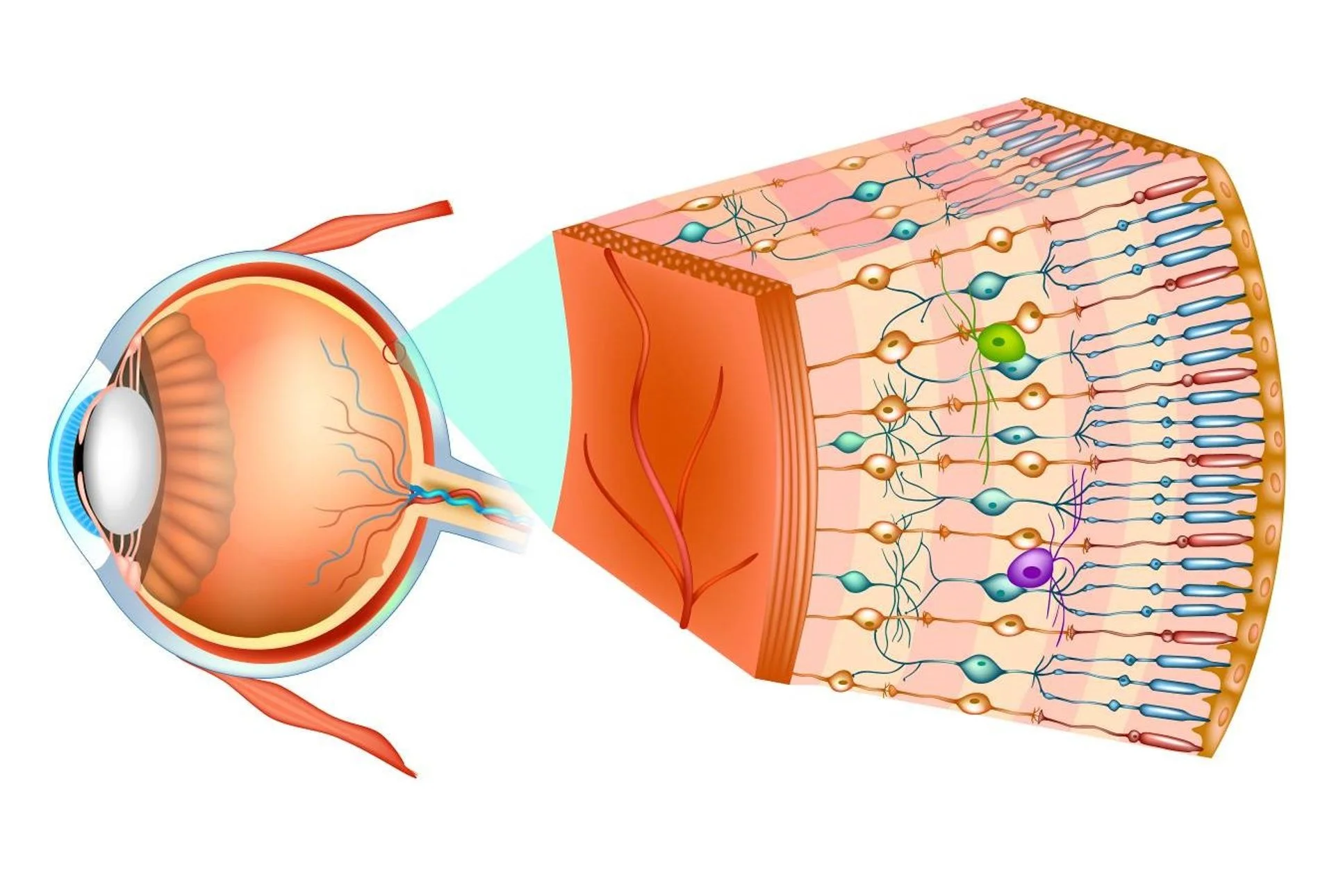
RETINA LIKE A “MINIATURE BRAIN”
The Professor explains: “Our work started several years ago from a collaboration between our Unit (directed by Prof. Francesco Bandello) and the Neurology Unit, directed by Prof. Giancarlo Comi”. Retina is a kind of “brain extroversion”, to which it remains connected through the optic nerve: “By studying the retina it is possible to make considerations about what is happening in the brain and study neurological pathologies such as multiple sclerosis, optic neuritis (inflammations of the optic nerve) and Alzheimer”.
The pathology we are telling about is focused precisely on the latter.
“Two populations have been seen at the Neurology Department: patients with established Alzheimer’s, and patients with MCI (Mild Cognitive Impairment), i.e. those who are not affected by Alzheimer’s yet, but who are at high risk of progression to the disease”.
These groups underwent a neurological evaluation that included several tests, such as electrocardiogram, electroencephalogram, tac or magnetic resonance, and biomarkers in the cerebrospinal fluid, especially the beta amyloid protein, a toxic substance involved in the development of Alzheimer’s, one of the most important biomarkers to make the disease’s diagnosis.
“Following this neurological visit, we took over ophthalmology consulting, visiting Alzheimer’s and MCI patients thanks to the tools we have integrated into our clinical practice:
OCT (optical coherence tomography) e OCTA (OCT angiography), two instruments similar to a CT scan, without using dangerous radiation, used to study the retinal structure and ocular vessels;
DVA (dynamic vessel analyzer), that studies the motility of blood vessels, donated by SOStegno 70, [a non-profit association born thanks to the support of the Childrens Endocrinology Center and IRCCS San Raffaele, which for years has been involved in the well-being of diabetic children and their families, Editor’s Note].
The study lasted about a year, after which we analyzed the data obtained from patients: the result was encouraging”.
ALZHEIMER’S AND MCI PATIENTS SHOW CELL LOSS IN THE RETINA
From a morphological point of view, the researchers have confirmed a data already present in the literature: “Overall we observed a cell loss in the retina, mainly located at the level of those layers more strictly in communication with the brain, i.e. the ganglion cells, which are expression of brain cells in the retina. In particular, it is the magnocellular cells, the larger ganglion cells (hence the name), which significantly decrease in number both in Alzheimer’s and MCI patients. Although MCI patients perform fairly normal daily activities, an important cell loss occurs in their retina (and in their brain), much like what happens in Alzheimer’s patients”.
SAME NUMBER OF BLOOD VESSELS, BUT LESS WORKING
The OCTA technique allowed researchers to obtain a new and unique data: “By analyzing blood vessels, we realized that Alzheimer’s and MCI patients have a vascular structure and a number of vessels similar to healthy subjects. We were very surprised by this result, considering that it contradicts what was recently reported by another study”. Thanks to the DVA instrument, they investigated vascular dynamics: “We found that in these patients the vessels are present in normal quantities, but they work poorly”. The device studies the vascular dynamics for 6 minutes, through 3 repeated cycles of stimulation to a particular light alternating with moments of rest: “Usually we observe a vasodilatation followed by a vasoconstriction, but in Alzheimer’s and MCI patients the vascular dynamics is altered, demonstrating a serious impairment of “neurovascular coupling” [the ability to increase the blood flow (and therefore the supply of oxygen and glucose) to neurons when they become more active, Editor’s Note]”.
Some members of the San Raffaele Hospital Ophtalmology Unit. Prof. Querques is in first row, in blue blazer; Dr. Borrelli is in first row, second from left. Prof. Bandello, Unit Director, is in the middle, back row.
LESS BETA AMYLOID IN THE CEREBROSPINAL FLUID
Says Dr. Enrico Borrelli, second author of the article and medical consultant of the structure: “This work has brought out another important result: in Alzheimer’s and MCI patients we recorded a close association between the decreased functioning of the retinal vessels and the decrease in beta-amyloid protein in the cerebrospinal fluid. Such decrease is due to a greater accumulation of this protein in the tissues, with consequent tissue damage. Other authors had already highlighted this correlation; the merit of our work is to have demonstrated it for the first time in vivo in the retina”.
Our study has shown that MCI patients already have all the typical characteristics of Alzheimer’s, although not necessarily a MCI patient will progress to dementia” clarifies Prof. Querques. How can we act on these patients to prevent or delay the damage caused by Alzheimer’s? “The neurologists must answer this question”, the Professor concludes. “Further studies will be needed, but our research opens the door to the hope that one day it may be possible to start early treatments to prevent Alzheimer’s: this is going to be a great challenge”.