Controfigure
Controfigure. Cells, tissues and their imitations
"Controfigure" is the photographic exhibition conceived and realized by Vita-Salute San Raffaele University and IRCCS San Raffaele Hospital of Milan.
The exhibition is based on a series of high-definition images (coloured and acquired with different microscopy techniques in the laboratories of San Raffaele) of organelles intracellular, cell and tissue of particular shape or appearance.
Each biological image is associated with a morphologically similar photograph (or object exposed as a sculpture), relating to the daily reality: such similarity is not only aesthetic, but also conceptual and functional.
"Controfigure" is a journey through colorful microscopy images and everyday objects, to discover that in our universe no idea (especially if particularly ingenious!) is wasted, which form and function are deeply connected to each other.
Shocks absorptions
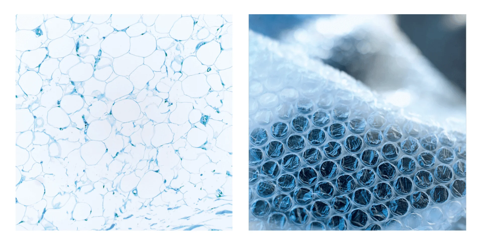
The bubble wrap is one of the most used packaging materials in the world: air bubbles trapped in the polyethylene sheet make it able to protect the most fragile objects from impact.
Our body contains a cushioning material too: the white adipose tissue, which is distributed throughout the body, particularly in the deep layers of the skin. White adipose tissue is an important energy reservoir, acts as a thermal insulator and protects organs from mechanical stress.
Just like bubble wrap, this tissue has a bubble-like appearance: its component cells, called adipocytes, are full of lipids (fats) that accumulate as small drops and gradually merge into a single voluminous drop that can take up all the available space. This is the reason why the components of the fat cell appear crushed under a microscope along their perimeter, while the lipid drop (which is dissolved by the reagents used in the staining process) appears as an empty space.
Iodized basins
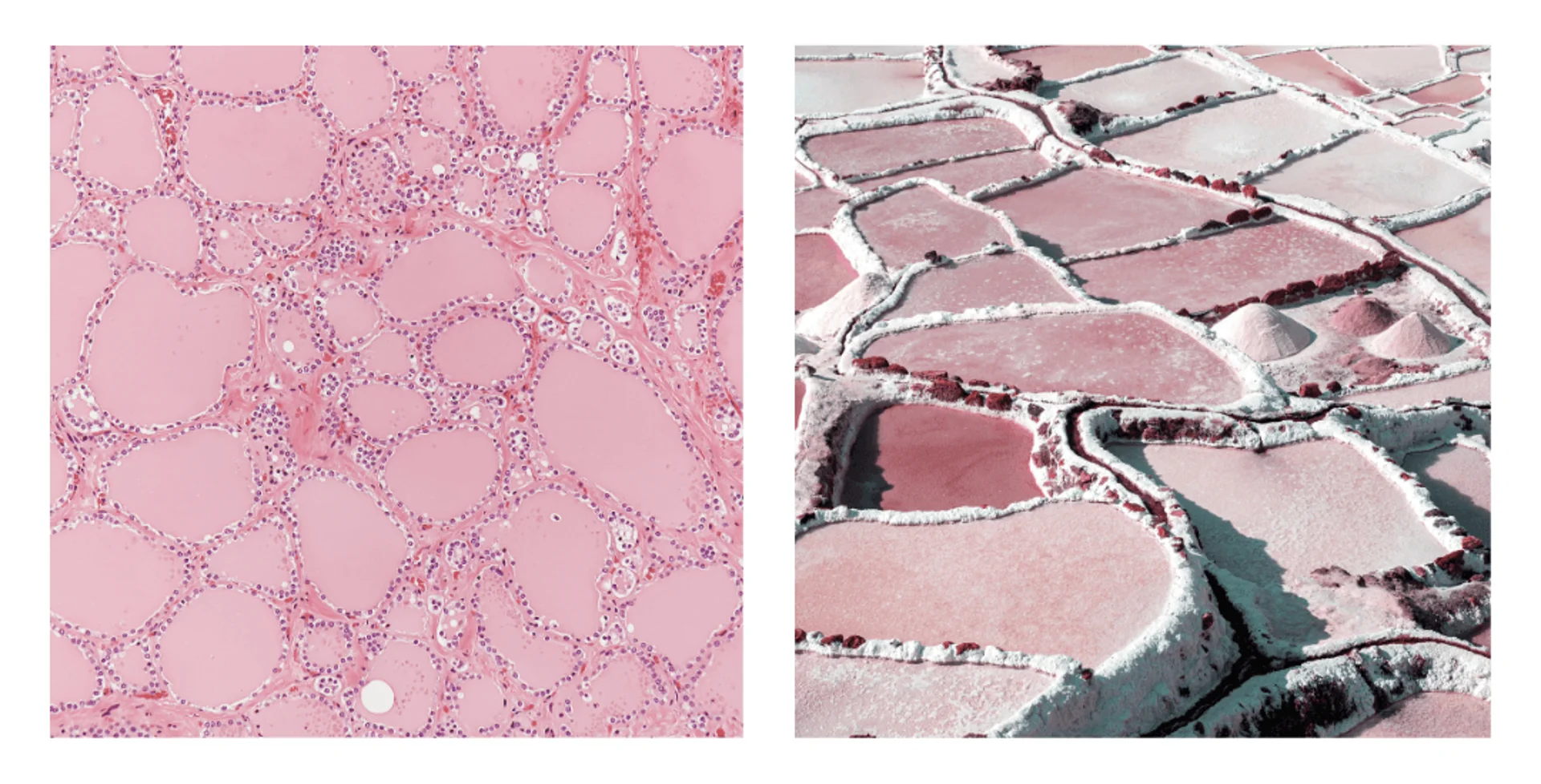
Saltworks are systems that extract salt from sea water through a passive process: water is collected in big artificial basins and it is left to evaporate in the sunlight. The salt that remains on the bottom of the basins contains iodine, a chemical element that is fundamental for the thyroid to function properly.
In fact, the thyroid follicles resemble saltworks. Each follicle is composed of a layer of secretory cells that surround an area of a tissue called colloid. Similarly to saltworks basins, iodine is stored in the colloid, as it is essential to synthesizing thyroid hormones, that regulate the metabolism and the development of many tissues in our body.
The iodine deficiency, an health problem of many developing countries, causes swelling of the thyroid and damage to the skin, hair, heart and cognitive functions. This is the reason why it is so important to consume iodized salt - which was put on the market precisely to reduce the risk of hypothyroidism.
Curiosity: a highly sought-after dye
In ancient times, blue was considered a symbol of authority and elegance. The natural raw material used to prepare blue dyes was a precious stone, lapis lazuli, which came from Afghanistan. In the 1500s, it was discovered that the indigenous people of the New World dyed their clothes an intense blue using extracts from a tree native to Central America: Haematoxylon campechianum.
Spain, which had established numerous colonies in the region, controlled the trade of this tree and was often targeted by English pirate galleons. From its valuable trunks, haematoxylin can be extracted, a colourless substance that oxidizes to become hematein, a dark purple colour tending towards blue. The use of haematoxylin as a dye for paints, lacquers, and inks persisted for a long time, even after the introduction of synthetic dyes in the 1800s, but its primary use today is in microscopy.
When used in combination with eosin, it allows for highlighting different components of the cell: haematoxylin binds to DNA and RNA present in the nucleus, colouring them blue-purple, while eosin binds to proteins in the cytoplasm, which appear pink.
A transport network
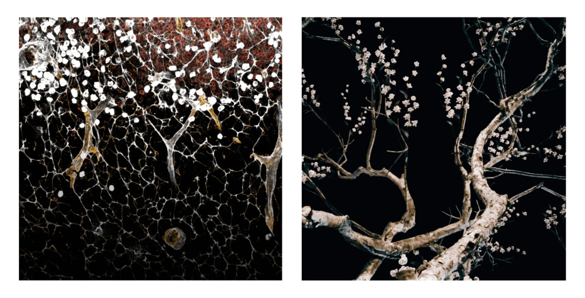
An important part of the immune response against viruses and foreign microorganisms is organized in filtering stations: the lymph nodes. In adults, there are approximately 600 to 800, distributed in several areas of the human body.
Each of these small organs contains a network of channels that carries the lymphatic fluid coming from tissues. If the lymph brings dangerous agents – viruses, bacteria or tumor cells – the B lymphocytes in the lymph nodes produce antibodies to fight them, and the same system of channels that receives the lymph is used to release these antibodies into the bloodstream.
Speaking of lymph, have you noticed that the channels of a lymph node resemble blossoming trees? Trees have a specialised vascular system: through this system the lymph is driven from the roots to every leaf, where photosynthesis happens, and the produced sugar molecules are distributed to all cells of the plant.
Electrical insulators
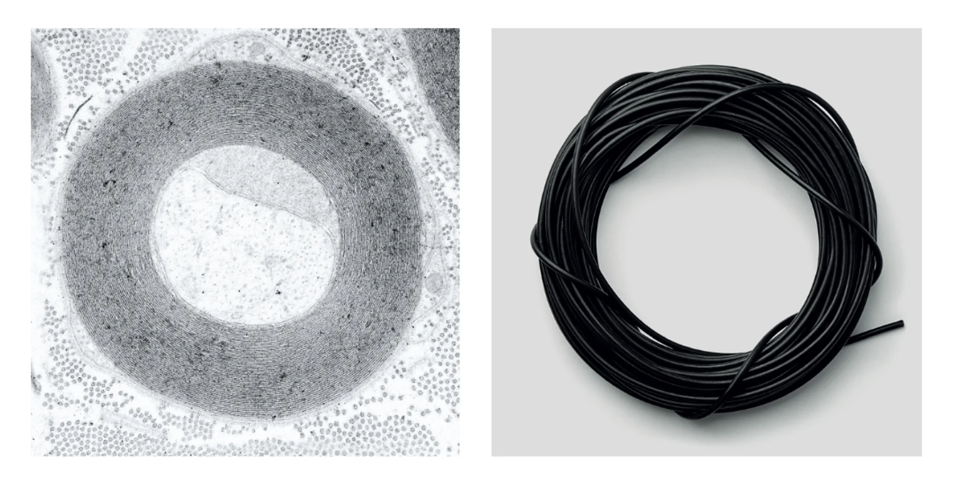
Do you know that neurons represent only 10% of our nervous system? The other 90% is composed of glial cells (from Latin “glia”, glue), that perform functions of structural support and metabolic functions. Among these there is the production of myelin, a substance fundamental to exchange nervous signals between neurons.
These signals need to travel long distances in a short time. For example, the sciatic nerve begins from the bottom of the spinal cord and arrives at the tip of the foot, about a metre away. To be sure that the impulse arrives at destination quickly and without dissipating during travel, nature has invented a smart system: the fibres are covered by an insulating sheath, called myelin.
This is such an efficient method that humans have been copying it – without knowing it – for over a century: the electric cables are made of a metal conductive covered by insulating materials, usually rubber, polyethylene or PVC.
Curiosity: gold-plated cells
In 1924, a young physicist, Louis de Broglie, had a great intuition: since light waves can behave like beams of particles (photons), then particles can behave and can be described as waves.
If this is true, illuminating an object is no longer the only way to make it visible. That is also possible by striking it with a beam of massive particles, such as electrons. This strategy has an important advantage: the high-frequency wave of electrons can detect objects at the nanometre scale!
Modern electron microscopes — like those used at San Raffaele in Milan — are extremely high-resolution instruments, capable of distinguishing the internal structures of a cell with extraordinary detail.
To protect the biological sample from the energetic impacts of electrons—which risk overheating and damaging it—and to improve image contrasts, researchers coat the tissue with a layer of metal. Which is the most used metal? Not exactly the cheapest: gold.
Hard bones
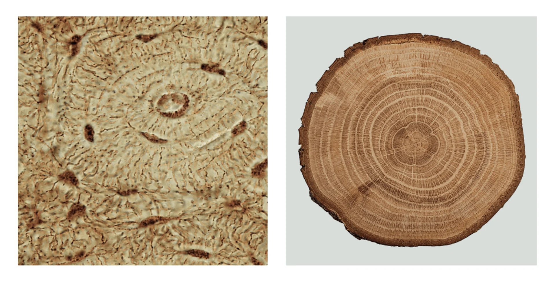
To support the weight of the entire body, to resist pressure, tension, traction… a lot of strength is needed! Thanks to their unique mineral composition (which includes crystals of calcium phosphate) and organic composition (which contains collagen fibres), bones are both resistant and flexible.
The base unit of the compact bone is the osteon: each osteon is composed of columns of concentric lamellae, like the growth rings of a tree, with a blood vessel in the centre. The structure – ideal to resist compressive forces – is constantly evolving different classes of cells maintain a balance between destroying old bone and laying down new bone, renewing approximately 10% of them each year.
Thanks to this dynamism the bones grow during youth, repair in case of a fracture, become more robust because of physical activities such as weightlifting or they become thinner instead in a low-gravity conditions, similarly to what happens to astronauts on the International Space Station (ISS).
Extracellular traps
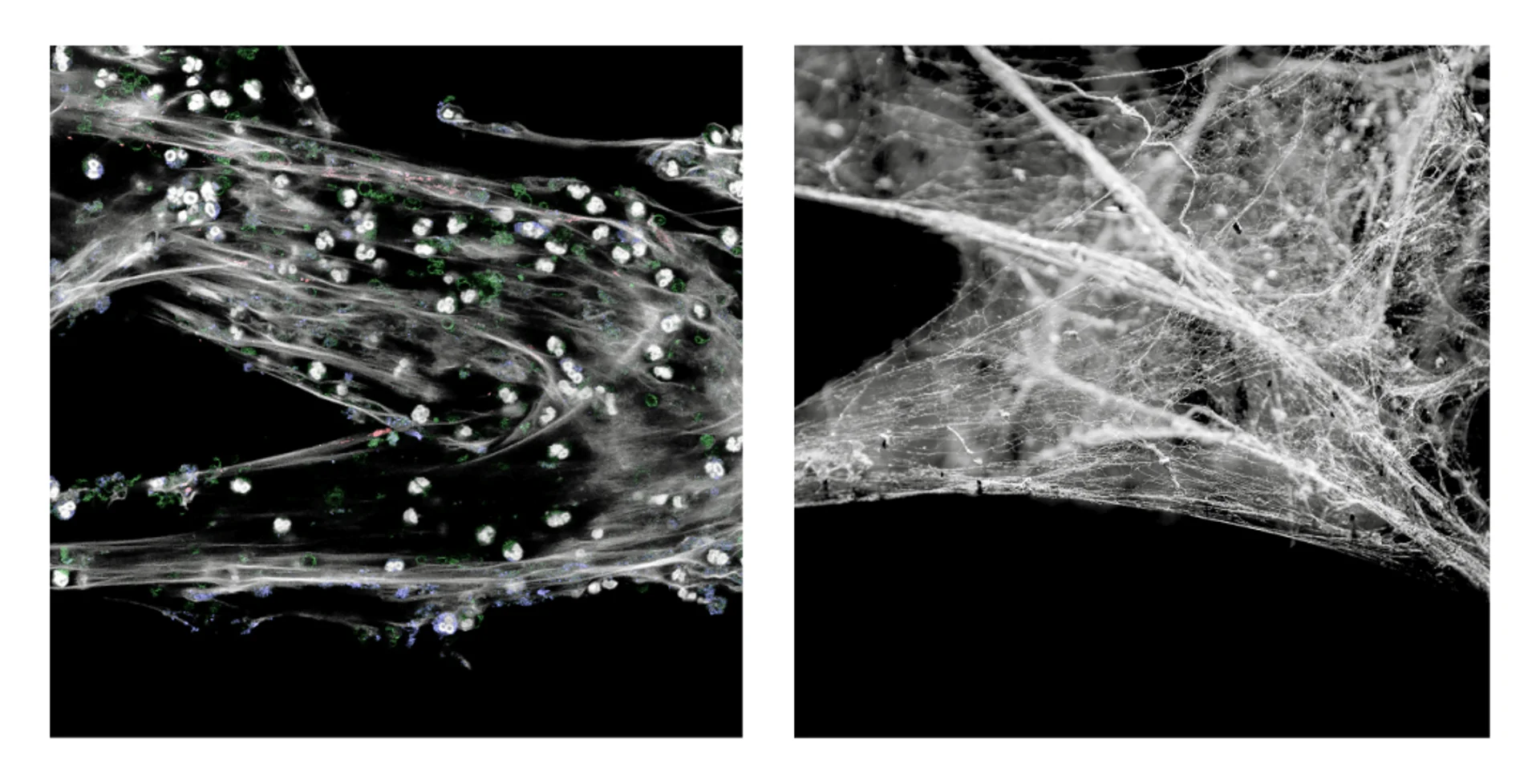
One of the stratagems used by the immune system to protect itself from attackers consists of sticky traps made by DNA fibres, proteins and enzymes. Similarly to a spider’s web, the dense net of filaments traps viruses and bacteria and neutralizes them.
Neutrophils, cells that constantly patrol our bodies travelling in the bloodstream and intervene in the early stages of an infection, are responsible for releasing the traps into the external environment. This is why the traps are called “Neutrophils Extracellular Traps”, NETs.
This strategy corresponds to a real suicide operation, which are made by neutrophils only in emergency situations, when there are too many pathogens to be phagocytised. The release of material usually hidden in the nucleus into the external environment is a signal for other immune system cells which immediately arrive and trigger an inflammatory reaction that can potentially be dangerous.
A microscopic archipelago
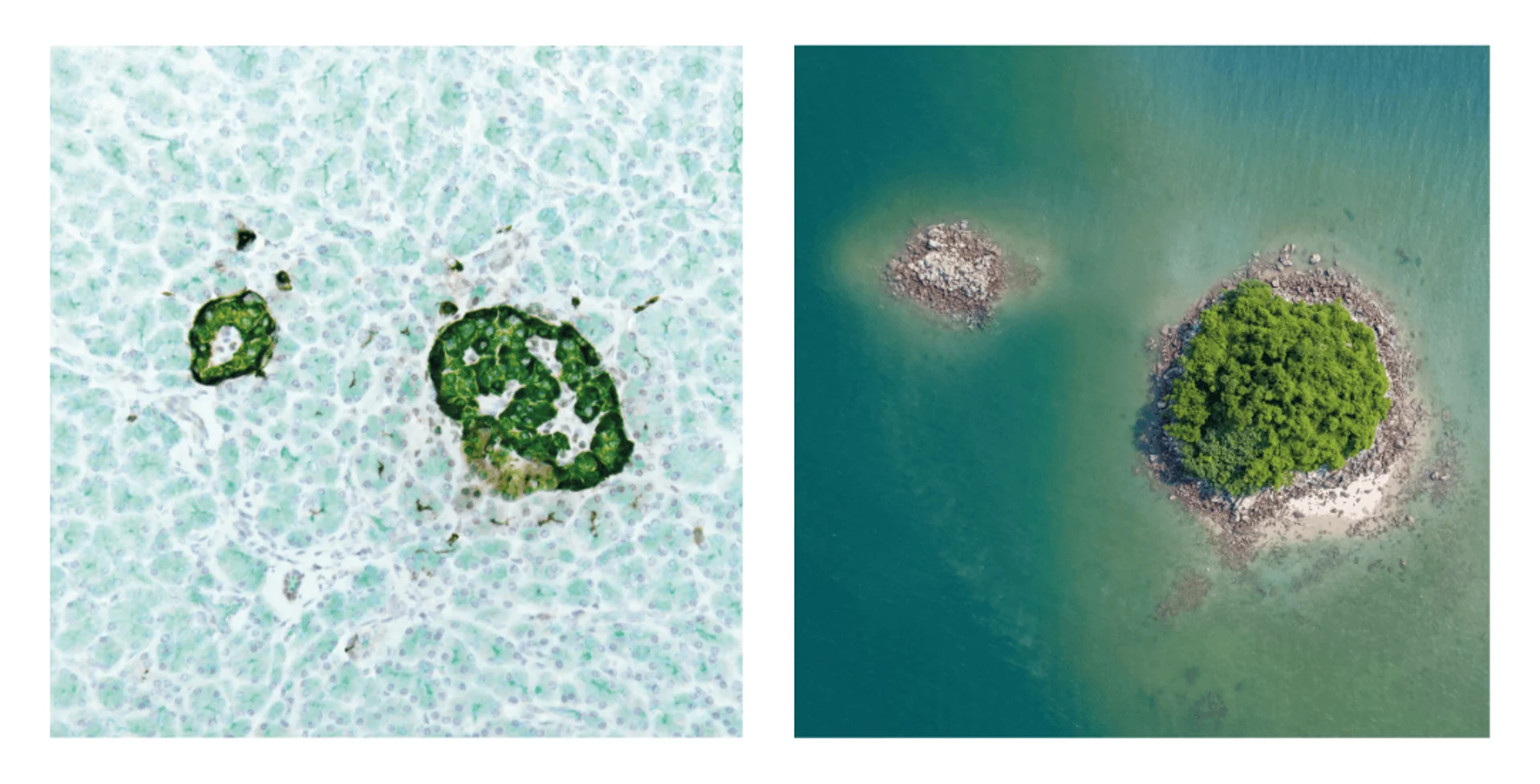
Did you know that the pancreas contains more islets than there are islands in a Pacific Ocean archipelago? They are called “Islets of Langerhans” , they are microscopic (their diameter varies from 0.3 to 0.7 mm) and each human pancreas contains more than one million of them.
The pancreatic islets are inhabited by different types of cells that are essential to regulate the metabolism of the entire organism. The best-known among them are the β (beta) cells: they are the only cells capable of producing insulin, the hormone that regulates the levels of sugar in the blood. In individuals with type-1 diabetes, the islets of Langerhans, destroyed by the immune system, can no longer supply insulin to the body.
In addition to β cells, these islands contain other cell populations, including α, δ, F and ε cells, which produce substances necessary to regulate digestion and nutrients absorption, as well as other functions that remain unknown to researchers studying these fascinating structures.
Geometric restrictions
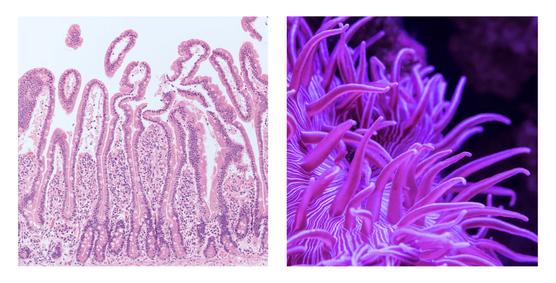
There is one geometric rule that makes life on planet Earth more difficult: when a three-dimensional object becomes larger, its surface area increases more slowly than its volume.
This implies that, proportionally, larger organisms – such as elephants or humans – have less surface area in contact with the external environment than smaller organisms, such as ants. This is a real problem considering that the exchange of heat, nutrients and oxygen with the environment happens through surfaces.
To solve this problem, in the most complex organisms the shape of many organs maximalizes the surfaces available. An example is given by the intestine, which hosts millions of villi: protuberances that are less than a millimetre long and whose thickness corresponds to that of a single layer of cells. Thanks to the villi the first part of the intestine (3-4 metres long) has a surface area equivalent to that of a tennis court!
Similar solutions can easily be found in nature, such as in the tentacular shape of sea anemones, which absorb nutrients from the surrounding water.
Cells like cities
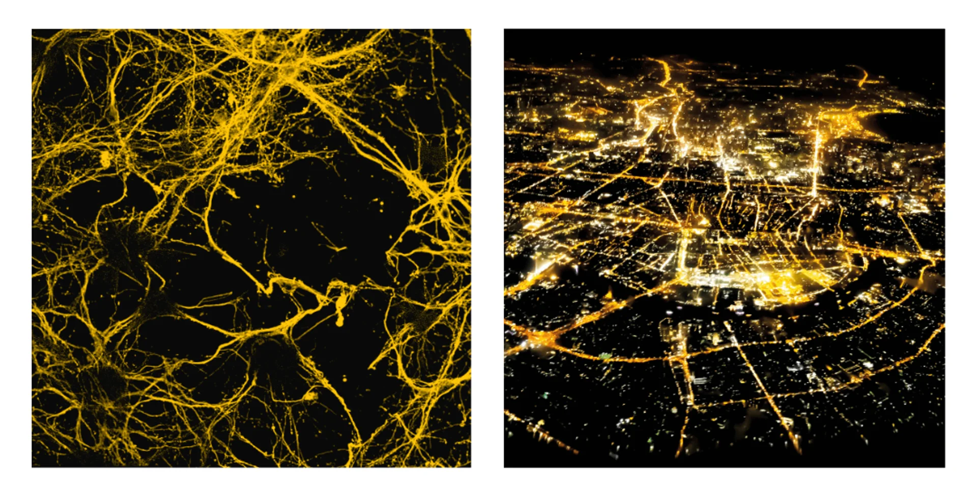
Maybe you have never noticed that, but neurons resemble cities viewed from above, as if seen from a taking-off airplane. The city centre reflects the cell body, while the streets that branch out towards the suburbs and connect the city with the neighbouring centres resemble axons and dendrites (extensions that allow neurons to receive and send signals to other nerve cells).
We shouldn’t be surprised by the resemblance between different networks, such as neural networks and road networks: both can be described using few parameters that don’t depend on the nature of the network but of its mathematical structure, such as the number of nodes or the frequency of connections.
Both are the result of local random processes – the construction of a street to connect a new district and the elongation of a dendrite toward a nearby cell – that produce order and complexity on a large scale.
It is this kind of order that allows us, in the first case, to write and read this text, in the second case to share space and resources with thousands – or millions – people.
A dynamic skeleton
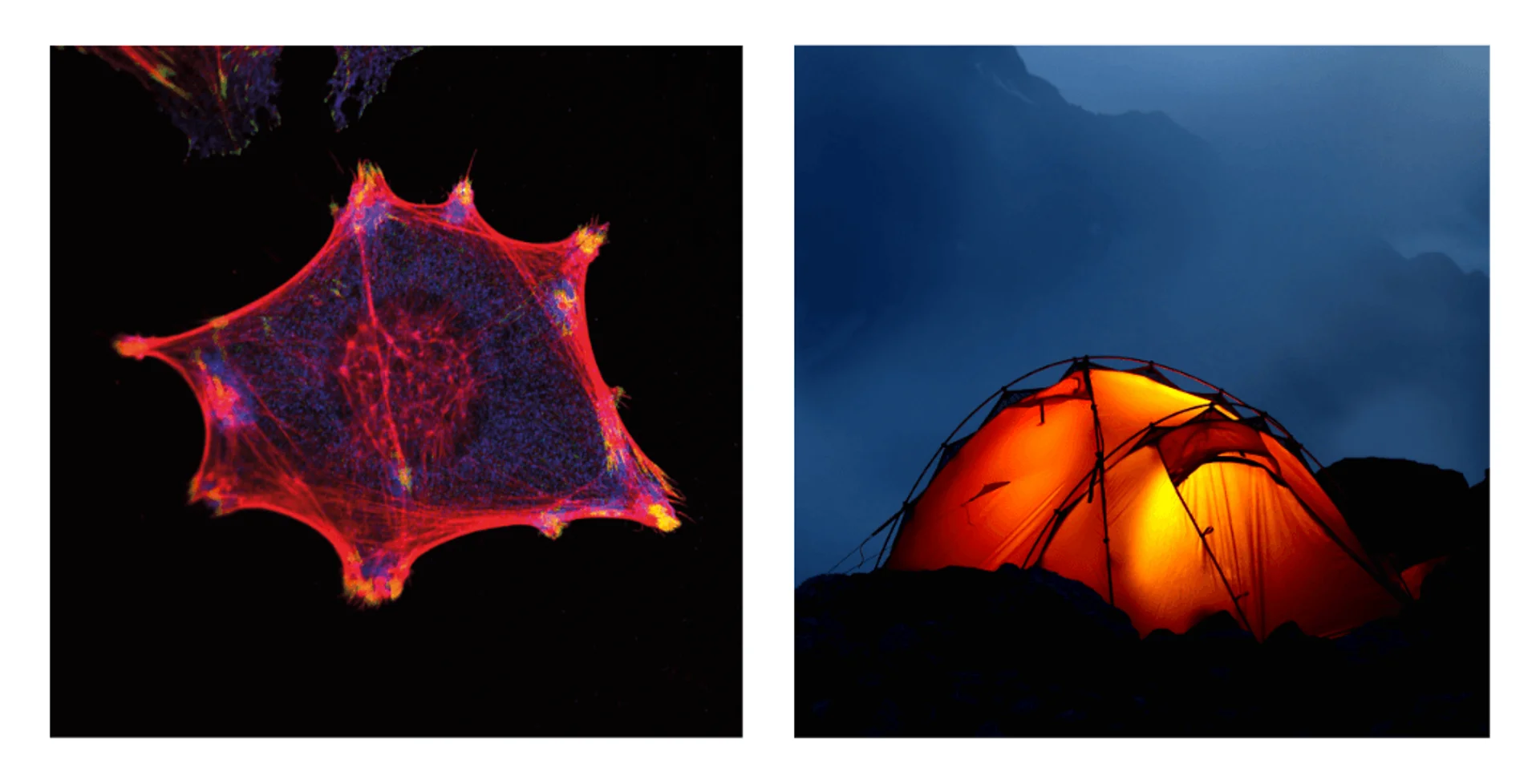
A membrane that acts as cloth and a framework that serves as support: it's all you need to define a cell as a real tensile structure.
The framework of a cell is the cytoskeleton (literally the "skeleton of the cell") which is composed of three fundamental elements: microtubules, microfilaments and intermediate filaments.
The organization of the various filaments in the cytoskeleton is an architectural masterpiece: microtubules push the membrane outward while microfilaments and intermediate filaments pull it inward creating a perfect dynamic balance.
This flexible organization - which responds to internal and external stimuli – allows for several essential functions: the cytoskeleton provides mechanical and structural support for the membrane and the organelles, facilitates intracellular transport of materials and enables the cell to adhere to other cells, contract and move.
Curiosity: nobel Prize jellyfish
In the Pacific Ocean lives the jellyfish Aequorea victoria: it is a few centimetres wide, carnivorous, and almost completely transparent. Nevertheless, in the ocean depths, it does not go unnoticed: the outer profile of its bell emits fluorescent green light, thanks to the presence of a special protein called GFP, Green Fluorescent Protein.
Its discovery revolutionized the world of microscopy, contributing to the awarding of the Nobel Prize in Chemistry in 2018: given its small size, GFP easily binds to other cellular structures without interfering with their activities, and when struck by ultraviolet light, it becomes fluorescent, allowing researchers to see under the microscope the structures it has bound to, otherwise indistinguishable from the rest of the cell.
Over the years, various versions have been created that emit fluorescent light in different colours: a rich palette that allows for the colouring (and viewing) of different parts of cells.