Thanks to an innovative imaging technology developed and tested at San Raffaele, physicians were able to improve diagnosis capacity from 50% to 90% in patients arriving at the emergency department for acute chest pain, allowing for more effective and timely intervention
Acute chest pain represents the first common cause of emergency department visits in the western world and it can be the main symptom of numerous pathologies, sometimes difficult to distinguish, especially in emergency. Chest pain can in fact be caused by a wide range of clinical conditions, from the most trivial ones, such as gastroesophageal reflux, to the most critical and dangerous, such as heart attack or acute myocardial inflammation. That is why identifying the cause underlying the pain quickly is of utmost importance in emergency medicine.
Today we are one step closer to successfully address the issue: doctors and researchers at San Raffaele research and university hospital in Milan have devised a new CT examination protocol that drastically improves, in both precision and speed, the diagnosis of the cause underlying acute chest pain. The procedure developed could soon become an international standard, as already suggested in the editorial that accompanied the publication in the prestigious peer-review journal Radiology.
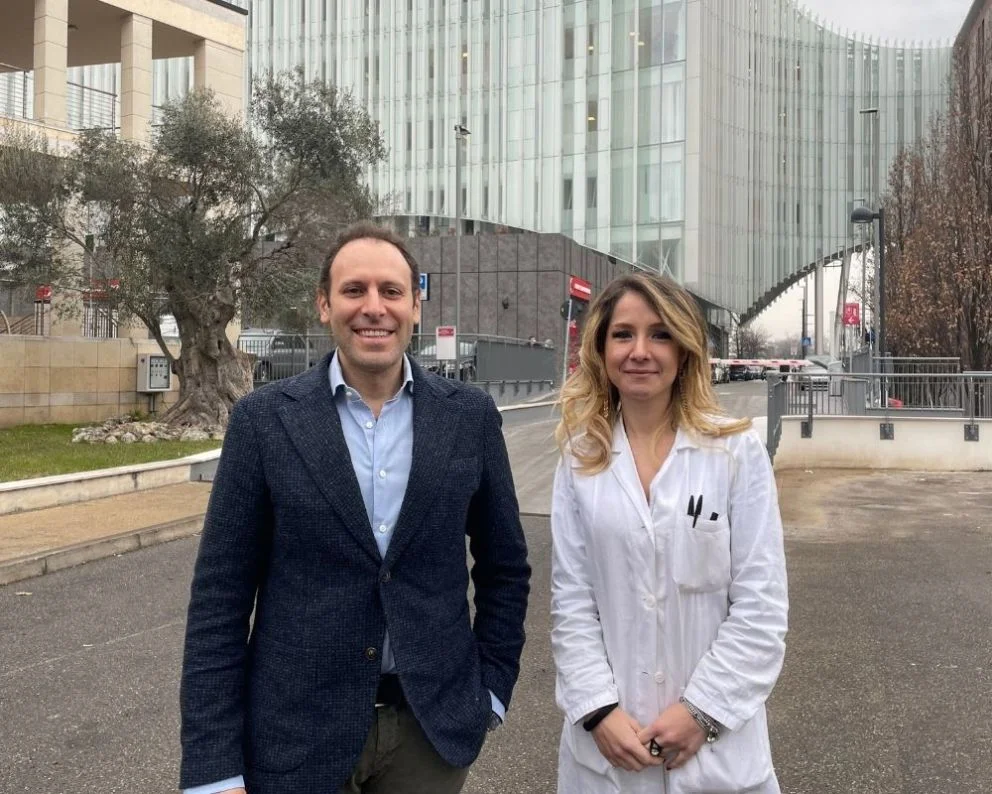
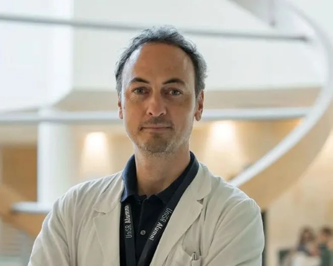
The research was coordinated by Antonio Esposito – full professor of Medical imaging and Radiotherapy at Vita-Salute San Raffaele University and deputy director of the Experimental Imaging Center of IRCCS Ospedale San Raffaele – and was possible thanks to the collaboration with the University of Turin and the Maria Vittoria hospital, also in Turin.
The challenge of diagnosis in patients with acute chest pain
On average, for every 100 emergency room visits, nine are due to sudden acute chest pain. The symptom, which can be a sign of severe conditions – such as heart attack, pulmonary embolism or myocarditis – is associated with a high risk of complications and death, especially in the case of inaccurate diagnosis and inappropriate discharge.
In the most advanced hospitals, patients with chest pain considered to be at intermediate risk after clinical evaluation undergo CT using an angiographic protocol (called angio-CT TRO). This protocol can exclude some dangerous pathological conditions, such as obstruction of the arteries that carry blood to the heart, potentially fatal pathologies of the first tract of the ascending aorta and pulmonary embolism. However, it also leaves unexplored a whole other series of equally dangerous conditions, which can produce lesions in heart muscle tissue and which must be treated in a timely manner.
"For this reason, unfortunately, even with the use of the Angio-TC TRO, the definitive diagnosis is reached in only 50% of patients. The other 50% remain without diagnostic confirmation, despite the presence of elevated troponin values in the blood, a biomarker indicating that heart cells are under intense stress”
explains Anna Palmisano, radiologist at IRCCS Ospedale San Raffaele and first author of the study published in Radiology, which Palmisano carried out during her doctoral thesis at Vita-Salute San Raffaele University.
“These patients, without a clear diagnosis, are usually kept under observation, monitored with repeated dosages of troponin, and subjected as soon as possible, in the following days and weeks, to further diagnostic investigations, such as a cardiac magnetic resonance or a biopsy”
continues Dr. Palmisano.
The consequence, however, is a delay in the diagnosis and in the discharge, with greater risk and stress for the patient, higher costs for the health system and greater crowding of the emergency departments and hospital wards. This is why the new diagnostic approach conceived at San Raffaele addresses a key unmet need in emergency health.
The new CT protocol will revolutionize the diagnosis of acute chest pain
The diagnostic protocol designed and tested at San Raffaele is grounded in two innovative imaging techniques, developed in the last ten years between Europe and the USA, able to add novel and fundamental information to the standard Angio-CT exam.
Both imaging techniques – called LCE CT and ECV CT – require a delayed CT scan that must be taken only 10 minutes after the classic angio-CT TRO and using the same radiocontrast infusion. Hence, they can be performed in the same session. San Raffaele is the first hospital to develop, test and use these techniques in the clinical context of acute chest pain, after being the first in the world, in 2016, to develop these same techniques for three-dimensional modeling and diagnosis of malignant ventricular arrhythmias.
“LCE CT and ECV CT make visible the alterations of the heart muscle tissue at the cellular and extracellular level, information previously inaccessible, allowing us to go beyond the evaluation of blood vessels and into new diagnostic possibilities. The combined protocol identifies both the presence of a damage in the tissue and its causes with extraordinary precision”
explains prof. Antonio Esposito, who coordinated the study "with enormous benefits for the patient and for the health system".
In the study published in Radiology, the protocol was tested on 84 patients with acute chest pain admitted to the ER: by combining the information provided by the two innovative techniques (LCE and ECV), without additional tests, it was possible to increase the diagnosis capacity from 50% to 90%. It means that for 42 of the 84 patients enrolled in the study, the new protocol was in fact crucial for diagnosing the pathology at the origin of chest pain.
“Of these 42 patients, 22 suffered from myocarditis, 3 from myocardial infarction with nonobstructive coronary arteries – not visible via CT angiography or coronary angiography – and fewer other specific myocardial diseases. All high-risk conditions that must be identified and adequately treated as soon as possible”
continues Esposito.
“This new CT could become the clinical standard in hospitals around the world, with an extraordinary impact on the management of patients with acute chest pain: anticipating diagnosis by days or weeks means saving lives and reducing healthcare costs at the same time."
You might be interested in
IgG4-Related Disease: A UniSR-Led Study on Obexelimab

Kidney Cancer: An AI Model Estimates Risk Before Surgery
Bone marrow transplant and leukaemia: understanding relapse mechanisms helps prevent them